Fetal Abnormalities Detection From Ultrasound Images Using Deep Learning
Introduction
Healthcare technology is rapidly evolving through the integration of Artificial Intelligence (AI), Machine Learning (ML) and Deep Learning. Medical image analysis is one of the important applications of these technologies, particularly in analyzing ultrasound images for prenatal assessment.
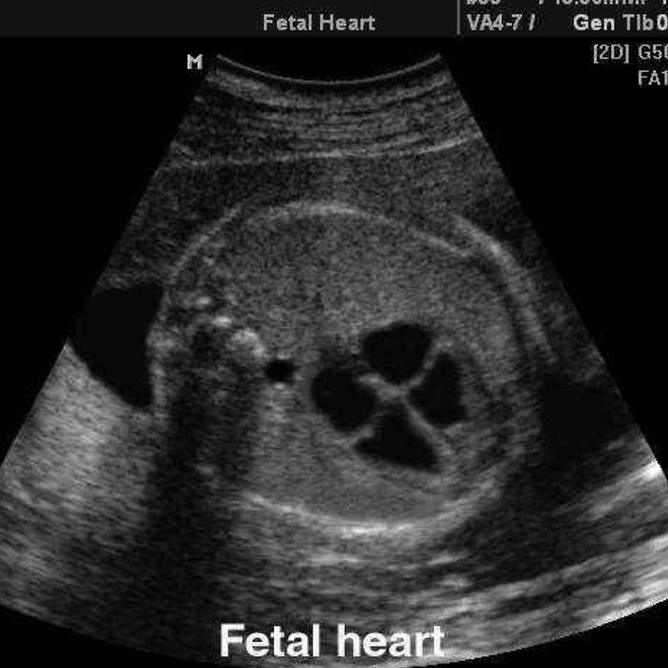
Ultrasound imaging is widely used during pregnancy to examine fetal growth, anatomical structures and development. However, interpreting ultrasound images requires specialized medical knowledge and experience. Variations in image quality, fetal position, scanning angles and image artifacts can make automated image analysis challenging.
The Fetal Abnormalities Detection From Ultrasound Images Using Deep Learning project explores an AI-based approach for analyzing fetal ultrasound images and identifying predefined abnormality patterns from a labeled research dataset.
The proposed system uses deep learning models to learn visual features from ultrasound images and classify them into predefined categories. Depending on the dataset and model design, the application can identify selected fetal anatomical patterns and distinguish between normal and abnormal image classes.
What Is Fetal Abnormality Detection Using Deep Learning?
Fetal abnormality detection using deep learning is a medical image analysis application that uses neural networks to recognize patterns in ultrasound images.
Convolutional Neural Networks (CNNs) are commonly used for image classification because they automatically learn important visual characteristics such as edges, textures, shapes and anatomical patterns.
The system processes an input ultrasound image, extracts relevant features and produces a classification result based on the categories included in the training dataset.
Transfer learning can also be used with pretrained architectures such as ResNet, EfficientNet and MobileNet to improve feature extraction and reduce training requirements.
Main Objectives
Develop a deep learning model for fetal ultrasound image classification.
Analyze ultrasound images using image preprocessing techniques.
Extract important visual features using CNN architectures.
Identify predefined normal and abnormal image categories.
Evaluate model performance using suitable classification metrics.
Provide visual predictions through a simple application interface.
Explore AI-assisted medical image analysis for research and educational purposes.
Importance of the Project in 2026
AI-assisted fetal ultrasound is an active research area. Recent research explores automated fetal biometry, anatomical segmentation, standard-plane recognition, image quality assessment and anomaly detection.
The 2026 research landscape also highlights explainable AI, vision transformers, federated learning and lightweight AI models for resource-constrained healthcare environments.
These developments make fetal ultrasound image analysis an interesting project area for students studying Artificial Intelligence, Deep Learning, Biomedical Engineering and Computer Vision.
Importantly, this project is an educational and research prototype. Its predictions should not be used to diagnose fetal conditions or make pregnancy-related medical decisions.
Key Features & Benefits
Working Principle of the Proposed System
The proposed system follows a deep learning-based image classification workflow to analyze fetal ultrasound images.
1. Ultrasound Image Dataset Collection
The first step involves collecting a labeled dataset containing fetal ultrasound images. Each image should have an appropriate reference label based on the dataset's documented classification criteria.
The dataset must be checked for image quality, duplicate samples, class distribution and labeling consistency.
2. Image Preprocessing
Ultrasound images may contain noise, speckle patterns, varying brightness and different resolutions.
Preprocessing operations can include:
Image resizing to a fixed input dimension.
Pixel normalization.
Noise reduction where appropriate.
Contrast enhancement.
Removal of irrelevant image borders when justified.
Conversion into the required image format.
3. Data Augmentation
Data augmentation increases training diversity by generating modified versions of existing images.
Possible augmentation techniques include rotation, horizontal or vertical transformations where anatomically appropriate, zooming, cropping and brightness adjustment.
Augmentation should preserve the anatomical meaning of the image and should only be applied to training data.
4. Deep Learning Feature Extraction
The preprocessed images are passed into a deep learning architecture.
A CNN learns different visual features through convolutional layers. Early layers identify basic patterns, while deeper layers learn more complex anatomical representations.
Popular architectures include:
Custom Convolutional Neural Network.
ResNet50.
EfficientNet.
MobileNet.
DenseNet.
Vision Transformer (ViT).
5. Classification Layer
The extracted features are passed to fully connected or classification layers.
A softmax output layer can estimate class probabilities for a multiclass classification task. For binary classification, sigmoid activation can be used.
The model assigns the input image to one of the categories defined in the dataset.
6. Model Training
The model is trained using labeled ultrasound images.
Common training components include categorical cross-entropy or binary cross-entropy loss, an optimizer such as Adam, and an appropriate learning rate.
Training and validation performance are monitored to identify overfitting and improve model generalization.
7. Prediction and Result Display
After training, a new ultrasound image can be supplied to the application.
The image is processed using the same preprocessing pipeline used during training. The trained model then produces a predicted class and its associated confidence score.
Suggested System Architecture
Ultrasound Image Input → Image Preprocessing → Data Normalization → CNN Feature Extraction → Deep Learning Classification → Prediction Evaluation → Result Visualization
Image Preprocessing Module: Resizes, normalizes and prepares input images.
Feature Extraction Module: Learns relevant image characteristics using deep learning.
Classification Module: Predicts the image category using the trained model.
Evaluation Module: Measures model performance using suitable evaluation metrics.
Visualization Module: Displays input images, predicted categories and model evaluation results.
Performance Evaluation Metrics
The model should be evaluated using more than accuracy, especially when the dataset contains unequal class distributions.
Accuracy: Overall percentage of correct predictions.
Precision: Proportion of positive predictions that are correct.
Recall (Sensitivity): Proportion of actual positive samples correctly identified.
Specificity: Proportion of actual negative samples correctly identified.
F1-score: Harmonic mean of precision and recall.
Confusion Matrix: Shows correct and incorrect predictions across classes.
ROC-AUC: Measures discrimination performance where appropriate.
For reliable evaluation, patient-level separation between training, validation and test sets is important to avoid data leakage.
Implementation Guide
Key Features of the Fetal Ultrasound Detection System
1. Automated Image Classification
The system analyzes ultrasound images and assigns them to predefined categories using a trained deep learning model.
2. CNN-Based Feature Extraction
Convolutional neural networks automatically learn important visual patterns from ultrasound images without requiring manual feature engineering.
3. Image Preprocessing
Image normalization, resizing and enhancement help prepare ultrasound images for consistent model input.
4. Transfer Learning Support
Pretrained deep learning architectures can be adapted to fetal ultrasound classification tasks using a suitable labeled dataset.
5. Prediction Confidence
The application can display model output probabilities to indicate how strongly the model favors a particular category. These values should not be interpreted as clinical diagnostic certainty.
6. Performance Visualization
Training accuracy, validation loss, confusion matrices and classification reports can be displayed to evaluate model behavior.
7. Expandable AI Architecture
The system can be extended with image segmentation, explainable AI and advanced deep learning models.
8. User-Friendly Interface
A web application can allow researchers and students to upload sample images and view experimental classification results.
Technologies Used
Component
Suggested Technology
Programming Language
Python
Deep Learning
TensorFlow / Keras
Image Processing
OpenCV
Numerical Processing
NumPy
Data Handling
Pandas
Visualization
Matplotlib
Machine Learning
Scikit-learn
CNN Architecture
ResNet / EfficientNet
Advanced Architecture
Vision Transformer
Backend
Flask / Streamlit
Development Tool
VS Code / Google Colab
The actual architecture should be selected according to the dataset size, image resolution and available computing resources.
Latest Trends in Medical Image AI – 2026
1. Vision Transformers
Vision Transformers use attention mechanisms to learn relationships between different image regions. They are being explored for complex medical image classification and segmentation tasks.
2. Explainable Artificial Intelligence
Explainable AI techniques such as Grad-CAM can generate visual heatmaps indicating which image regions influenced a model's prediction. These visualizations can help researchers investigate model behavior but do not establish that a prediction is medically correct.
3. Automated Ultrasound Image Segmentation
Deep learning segmentation models can identify anatomical structures such as the fetal head, abdomen and other regions of interest.
4. Federated Learning
Federated learning enables collaborative model development across participating institutions while keeping source medical data within local systems, subject to appropriate privacy and security controls.
5. Lightweight Deep Learning
MobileNet and optimized neural networks can help reduce computational requirements for research applications running on limited hardware.
6. Multimodal Medical AI
Emerging research explores combining ultrasound images with structured clinical measurements and other relevant information to develop more comprehensive analytical systems.
Recent 2026 reviews discuss these opportunities alongside limitations involving dataset diversity, external validation, clinical workflow integration and algorithmic bias.
Real-World Research Applications
Automated fetal ultrasound image analysis.
Fetal anatomical structure recognition.
Ultrasound image quality assessment.
Fetal biometric measurement research.
Medical image segmentation.
AI-assisted prenatal imaging research.
Clinical decision-support research.
These are broader research applications of medical imaging AI and should not be assumed to be fully implemented in every version of this student project.
Technical Specifications
Implementation Steps
The fetal abnormality detection project can be developed using Python and deep learning frameworks through the following stages.
Step 1: Requirement Analysis
Identify the project objectives, ultrasound image categories, dataset requirements and expected classification outputs.
Step 2: Dataset Collection
Collect suitable fetal ultrasound images from documented research datasets. Verify image labels, data quality and class distribution before training.
Step 3: Dataset Preparation
Organize the images into training, validation and testing datasets. Ensure that images from the same patient do not appear across different dataset splits.
Step 4: Image Preprocessing
Resize the images, normalize pixel values and apply suitable preprocessing operations to improve input consistency.
Step 5: Data Augmentation
Apply appropriate transformations to increase the diversity of training samples and reduce overfitting.
Step 6: Deep Learning Model Development
Build a CNN architecture or use transfer learning with ResNet50, EfficientNet or MobileNet to extract important ultrasound image features.
Step 7: Model Training
Train the model using labeled images and monitor training loss, validation loss and classification performance.
Step 8: Model Evaluation
Evaluate the trained model using accuracy, precision, recall, specificity, F1-score and confusion matrix analysis.
Step 9: Application Development
Develop a simple interface using Flask or Streamlit to upload ultrasound images and display experimental classification results.
Step 10: Testing and Validation
Test the system using an independent test dataset and analyze incorrect predictions, class-wise performance and model limitations.
Advantages of the Proposed System
Automates image classification for predefined research categories.
Reduces repetitive manual image labeling tasks during experimentation.
Uses CNNs for automatic feature extraction.
Supports transfer learning with pretrained architectures.
Provides measurable model evaluation results.
Enables visualization of training and prediction outputs.
Can be extended with explainable AI techniques.
Provides practical experience in AI-based medical image analysis.
Limitations and Challenges
Ultrasound images can contain noise and imaging artifacts.
Limited datasets may result in overfitting.
Differences between ultrasound devices can affect model performance.
Incorrect or inconsistent labels can affect training quality.
Class imbalance may reduce minority-class detection performance.
A model trained on one dataset may not generalize to other hospitals or populations.
High-quality clinical validation requires substantial data and expert oversight.
Future Scope
The proposed project can be enhanced by incorporating advanced deep learning architectures such as Vision Transformers and hybrid CNN-transformer models.
Future versions can integrate segmentation networks such as U-Net to identify specific anatomical regions before classification.
Explainable AI techniques can be added to visualize the regions that contribute to model predictions. Federated learning may also support research across multiple institutions without centralizing all patient images.
The long-term research direction is to develop reliable, transparent and carefully validated AI systems that may support qualified healthcare professionals in medical image assessment.
Conclusion & Next Steps
Conclusion
The Fetal Abnormalities Detection From Ultrasound Images Using Deep Learning project demonstrates the application of artificial intelligence and computer vision in medical image analysis.
By combining ultrasound image preprocessing, CNN-based feature extraction, deep learning classification and model evaluation, the proposed system provides an experimental approach for identifying predefined image categories.
The project introduces students to important concepts such as convolutional neural networks, transfer learning, image augmentation, classification metrics and explainable AI.
It also provides an opportunity to explore advanced medical imaging technologies, including Vision Transformers, image segmentation and federated learning.
Although deep learning offers promising research possibilities, the model's performance must be evaluated using independent datasets and appropriate validation procedures. The project is intended for academic and research purposes and is not a substitute for professional fetal assessment, prenatal screening or medical diagnosis.
Aislyn Technologies – Bangalore
Looking for expert guidance to develop your AI, Deep Learning or final-year project?
Aislyn Technologies provides technical guidance and project development support in Artificial Intelligence, Machine Learning, Deep Learning, Computer Vision, Medical Image Processing and Full-Stack Development.
Phone: +91 97395 94609
Email: info@aislyntech.com
Website: https://aislyn.in
Contact us today to start building your AI and Deep Learning project in Bangalore with our expert support!