Introduction to Lung Disease Prediction Using Deep Learning
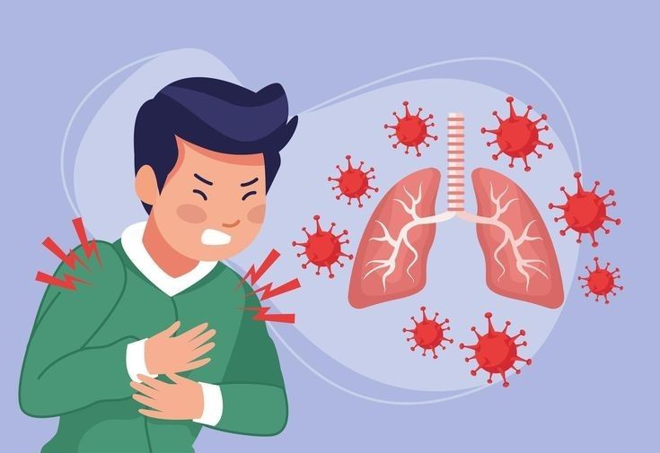
Lung diseases such as pneumonia, tuberculosis and other respiratory conditions can affect breathing and overall health. Medical imaging techniques, including chest X-rays and computed tomography (CT) scans, are widely used by healthcare professionals to examine lung structures and identify abnormalities.
Artificial intelligence and deep learning are creating new opportunities in medical image analysis. Convolutional Neural Networks (CNNs) can learn visual patterns from medical images and classify them into predefined categories.
Lung Disease Prediction from X-Ray and CT Scan Images Using Deep Learning is an AI-based project designed to analyse medical images and identify patterns associated with different lung conditions.
The proposed system combines image preprocessing, feature extraction and deep learning classification to generate automated prediction results. It aims to support medical image research and assist healthcare professionals in reviewing imaging data.
Key Features & Benefits
Objectives of the Proposed System
Analyse chest X-ray and CT scan images using deep learning.
Identify visual patterns associated with lung abnormalities.
Apply image preprocessing techniques to improve input quality.
Develop CNN-based image classification models.
Classify images into predefined lung disease categories.
Display prediction results through a user-friendly interface.
Evaluate model performance using standard classification metrics.
Support AI-based medical imaging research.
Implementation Guide
Technologies and Deep Learning Algorithms
Technologies Used:
Python: Main programming language.
TensorFlow and Keras: Deep learning model development.
OpenCV: Medical image preprocessing.
NumPy and Pandas: Dataset preparation and numerical processing.
Scikit-learn: Model evaluation and performance analysis.
Flask or Streamlit: Web-based application development.
Matplotlib: Image visualization and training analysis.
Deep Learning Algorithms
Convolutional Neural Network (CNN): Extracts important visual features from X-ray and CT images for classification.
ResNet: Uses residual connections to learn complex image features through deeper neural network architectures.
VGG: Provides a convolution-based architecture for medical image classification experiments.
EfficientNet: Offers an efficient architecture for extracting image features with optimized computational requirements.
Transfer Learning: Uses pretrained image models and fine-tunes them for lung image classification tasks.
Technical Specifications
Working Process of the Lung Disease Prediction System
Step 1: Medical Image Collection
Collect labelled chest X-ray and CT scan images from suitable medical imaging datasets.
Step 2: Image Preprocessing
Resize images, normalize pixel values and prepare the dataset for model training and prediction.
Step 3: Dataset Augmentation
Apply suitable transformations such as rotation, flipping and scaling to increase training data diversity.
Step 4: Deep Learning Model Training
Train the CNN or selected pretrained architecture using labelled medical images.
Step 5: Feature Extraction
The trained model learns visual characteristics such as lung patterns, textures and image structures.
Step 6: Disease Classification
Upload a new image and process it through the trained model to generate a prediction for the supported categories.
Step 7: Result Visualization
Display the predicted category and confidence score through a web application dashboard.
Step 8: Model Evaluation
Evaluate the system using accuracy, precision, recall, F1-score and confusion matrix.
Conclusion & Next Steps
Applications and Advantages
Applications
AI-based medical image analysis research.
Chest X-ray classification.
CT scan image classification.
Healthcare deep learning projects.
Medical image processing applications.
Academic research in artificial intelligence.
Advantages
Automated medical image classification.
Deep learning-based visual feature extraction.
Supports analysis of multiple image types.
Reduces repetitive manual image sorting.
Provides a user-friendly prediction interface.
Supports experimentation with advanced CNN architectures.
Future Enhancements
The system can be enhanced with explainable AI techniques such as Grad-CAM to highlight image regions influencing model predictions. Other improvements include multi-disease classification, lung segmentation, larger medical datasets, cloud-based image analysis and integration with medical imaging platforms.
Conclusion
Deep learning provides promising opportunities for analysing chest X-ray and CT scan images. By combining image preprocessing, CNN architectures and transfer learning, this project explores an automated approach to lung image classification.
The proposed system can support medical imaging research and AI-assisted image review. Its predictions are not medical diagnoses and must not replace radiologist interpretation, clinical examination or professional medical advice.
Contact Details
Aislyn Technologies, Bangalore
Phone: +91 97395 94609
Email: info@aislyntech.com
Website: https://aislyn.in
Contact us today to start building your Embedded project in Bangalore with our expert support!