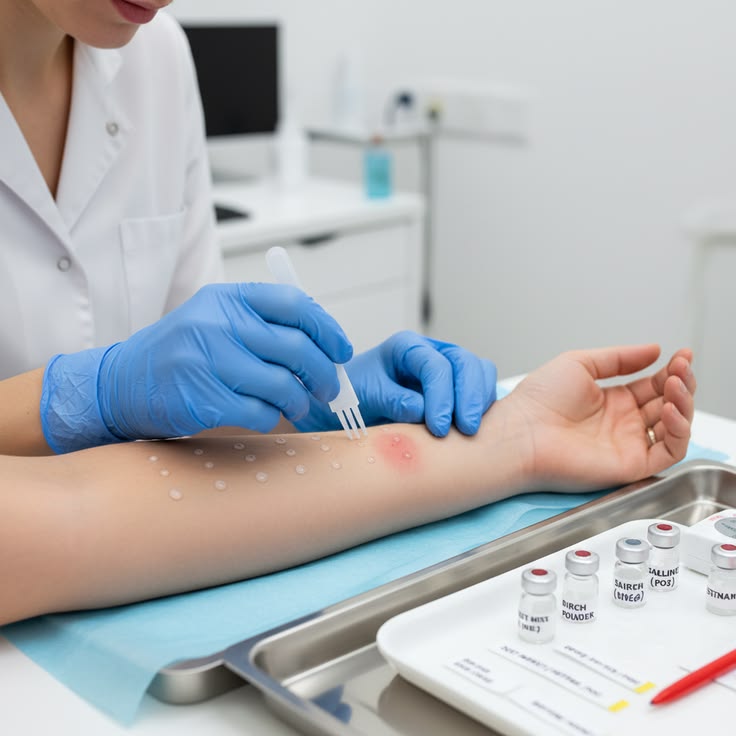
Skin prick testing is a widely used clinical procedure to help identify potential allergic reactions. During this test, a small amount of an allergen is introduced into the superficial layer of the skin, and the resulting reaction is evaluated by observing the wheal, a raised area of skin that develops at the test location. Accurate identification and measurement of the wheal are important for consistent interpretation of test results.
Traditional wheal assessment often involves manual observation and measurement, which can be affected by image quality, lighting conditions, skin texture and differences between observers. These challenges create opportunities for computer vision and artificial intelligence to support more consistent image analysis.
The Skin Prick Test Wheal Detection in 3D Images Using Convolutional Neural Networks project proposes a deep learning-based approach to identify wheal regions from three-dimensional skin images. By analysing surface information and image features, the system aims to detect the raised reaction area and estimate its dimensions.
This BCA project demonstrates how 3D image processing, CNN architectures and automated measurement techniques can be combined for medical image analysis. It is designed as a research and educational prototype rather than a replacement for clinical assessment.
Key Features & Benefits
Existing System and Its Challenges
In conventional skin prick test assessment, healthcare professionals examine the skin reaction and manually measure the wheal using appropriate clinical measurement methods. Although this approach is commonly used, manual assessment can introduce variations.
Some challenges associated with traditional assessment include:
Observer variation: Different professionals may identify or measure the wheal boundaries differently.
Irregular wheal shapes: Skin reactions may not have perfectly circular boundaries, making measurement more difficult.
Image quality: Lighting, shadows, skin texture and camera positioning can affect visual interpretation.
Time-consuming assessment: Repeated manual examination and measurement may require additional effort.
Limited digital documentation: Manual measurements may not provide consistent image-based records for later comparison.
Computer vision can help address some of these challenges by processing digital images and identifying regions of interest. However, reliable automated analysis requires suitable image acquisition, representative training data and careful validation.
Implementation Guide
Proposed System and Methodology
The proposed system uses a Convolutional Neural Network (CNN) to analyse 3D skin images and identify possible wheal regions. It combines image preprocessing, feature extraction, region detection and measurement estimation.
The methodology consists of the following stages:
1. 3D Image Acquisition
Three-dimensional skin images are collected using a suitable imaging device or prepared dataset. The images should capture sufficient surface detail to distinguish raised wheal regions from surrounding skin.
2. Image Preprocessing
The input images are prepared for model analysis. Preprocessing may include resizing, normalization, noise reduction, image alignment and removal of irrelevant background regions.
3. CNN-Based Feature Extraction
A CNN learns important visual and spatial patterns from the input images. Features such as surface boundaries, texture differences and wheal-like structures can help the model distinguish the reaction region from normal skin.
4. Wheal Region Detection
The trained model identifies the suspected wheal area. Depending on the selected architecture, this stage can use image classification, object detection or segmentation to locate the region.
5. 3D Measurement Estimation
After identifying the wheal region, the system uses the available 3D spatial information to estimate relevant dimensions, such as width, height and surface elevation. Accurate physical measurements require appropriate camera calibration, scale information and 3D reconstruction.
6. Result Visualization
The detected region can be displayed on the original image or as a 3D visualization. The system can present estimated measurements and save the analysis results for further review.
Technical Specifications
Technologies, Features and Applications
The project can be implemented using Python and deep learning frameworks, together with suitable 3D image processing libraries.
Technologies Used
Python – Main programming language.
TensorFlow or PyTorch – CNN model development and training.
OpenCV – Image preprocessing and computer vision operations.
NumPy – Numerical operations and image data handling.
Matplotlib – Image visualization and analysis graphs.
Scikit-learn – Model evaluation and performance analysis.
3D image processing libraries – Handling and visualization of volumetric or surface data.
Streamlit or Flask – Optional interface for uploading images and viewing results.
Key Features
3D skin image input and preprocessing.
CNN-based feature extraction.
Automated identification of suspected wheal regions.
Wheal boundary visualization.
Estimation of wheal dimensions from calibrated 3D data.
Storage and display of analysis results.
Model evaluation using suitable detection or segmentation metrics.
Potential Applications
Research in automated allergy-test image analysis.
Medical image processing education.
Development of computer-assisted wheal measurement prototypes.
Comparative analysis of image-based measurement techniques.
Research into deep learning for skin surface analysis.
The system's outputs should be treated as research measurements. Clinical use would require validation against accepted measurement methods, representative clinical datasets and review by qualified healthcare professionals.
Conclusion & Next Steps
Conclusion
Skin Prick Test Wheal Detection in 3D Images Using Convolutional Neural Networks demonstrates the potential of artificial intelligence and 3D computer vision in medical image analysis. By combining image preprocessing, CNN-based feature extraction, wheal region detection and 3D measurement estimation, the project provides an educational prototype for exploring automated skin reaction analysis.
This project is suitable for BCA students interested in deep learning, Python programming, computer vision and healthcare technology. It also provides a foundation for further research into accurate and reliable medical image analysis systems.
Contact Details
Aislyn Technologies, Bangalore
Phone: +91 97395 94609
Email: info@aislyntech.com
Website: https://aislyn.in
Contact Aislyn Technologies for project development, implementation guidance and technical support. Our team can help you develop your BCA final-year project with expert support.