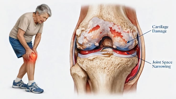
Osteoarthritis is a common joint condition that affects cartilage and surrounding bone, particularly in the knees, hips and hands. Early identification can help healthcare professionals assess joint changes and plan appropriate care. However, manually examining medical images can require considerable time and specialist expertise.
The AI-Based Osteoarthritis Detection Using Deep Learning project explores how artificial intelligence and medical image processing can support the analysis of X-ray images. By applying deep learning techniques, the system can learn visual patterns associated with different stages of osteoarthritis and generate predictions from uploaded images.
This Python-based project demonstrates the growing role of AI in medical image analysis and healthcare research.
Key Features & Benefits
How the System Works
The proposed system follows a structured image-processing and prediction workflow:
Image Upload: The user uploads a knee X-ray image through the application.
Image Preprocessing: The image is resized, normalized and prepared for model analysis.
Feature Learning: A deep learning model learns important visual patterns from the training images.
Classification: The trained model predicts the corresponding osteoarthritis category.
Result Display: The application displays the predicted category for review.
The quality of predictions depends on the dataset, image quality, model architecture and training process.
Implementation Guide
Technologies and Deep Learning Algorithms
The project can be developed using Python and popular AI libraries for medical image processing.
Software Technologies:
Python
TensorFlow or Keras
OpenCV
NumPy and Pandas
Scikit-learn
Flask or Streamlit
Possible Deep Learning Models:
Convolutional Neural Network (CNN)
VGG16
ResNet
EfficientNet
Transfer Learning
CNN-based models are particularly useful for learning spatial patterns from images. Transfer learning can also help reuse features learned from previously trained image-classification models.
Technical Specifications
Applications and Benefits
AI-assisted osteoarthritis image analysis has potential applications in medical imaging research and healthcare technology development.
Supports automated analysis of knee X-ray images.
Helps identify image patterns associated with osteoarthritis.
Demonstrates deep learning applications in medical image classification.
Provides a foundation for research into computer-aided assessment.
Helps students understand Python, image processing and neural networks.
Can be extended with model evaluation, visualization and reporting features.
The project can also be expanded to compare multiple deep learning architectures and evaluate their performance using accuracy, precision, recall, F1-score and confusion matrices.
Conclusion & Next Steps
Conclusion
The Revolutionizing Osteoarthritis Detection Using Deep Learning project demonstrates how artificial intelligence can be applied to medical image analysis. By combining Python, image preprocessing and deep learning models, the application can classify X-ray images into categories learned from its training dataset.
This project is suitable for students exploring AI, machine learning, deep learning and image processing. It can also serve as a starting point for further research into automated medical image analysis.
Important: This application is intended for academic and research purposes. Its predictions should not be treated as a medical diagnosis or used as a replacement for evaluation by qualified healthcare professionals.