ABSTRACT:
In today’s modern medical arena patients with brain tumor are increasing rapidly with a fast pace and above the par. Detection of brain tumor has become a challenging task to compete with. In this paper an automated method for detecting brain abnormalities and tumor edema has been proposed using sobel edge detection method. Various MRI images have been used as inputs here. Here, first of all the pre-processing of image has been done to cut out any discrepancy in it and then the image has been smoothened using median filter. We have proposed an appropriate method to find threshold value using standard deviation and we get an intensity map. Now we recomputed standard deviation for this intensity map. Using this we will calculate an average intensity of the pixels those are above this standard deviation. Finally, this computed average intensity will be taken as the threshold value to segment the tumor from the original MRI images. The intensity value greater than and equal to the calculated threshold value is set to 255 and less than is set to 0, this segments our abnormal region which is tumor. At last, we use sobel edge detector to identify the border of the tumor region. The outcome of the proposed method improves efficacy and accuracy for detection of brain tumors.
INTRODUCTION:
A brain tumor occurs when abnormal cells form within the brain. There are two main types of tumors: cancerous (malignant) tumors and benign (non-cancerous) tumors. Cancerous tumors can be divided into primary tumors, which start within the brain, and secondary tumors, which have spread from elsewhere, known as brain metastasis tumors. All types of brain tumors may produce symptoms that vary depending on the part of the brain involved. These symptoms may include headaches, seizures, problems with vision, vomiting and mental changes. The headache is classically worse in the morning and goes away with vomiting. Other symptoms may include difficulty walking, speaking or with sensations. As the disease progresses, unconsciousness may occur.
In brain tumor diagnosis, doctors integrate their medical knowledge and brain magnetic resonance imaging (MRI) scans to obtain the nature and pathological characteristics of brain tumors and to decide on treatment options. However, in brain MRI, where a great number of MRI scans taken for every patient, physically detecting and segmenting brain tumors is monotonous. Therefore, there is a need for computer aided brain tumor detection and segmentation from brain MR images to overcome the problems involved in the manual segmentation. Number of methods has been proposed in recent years to seal this break, but still there is no generally customary automated technique by doctors to be used in clinical floor due to accuracy and robustness issues.
In recent times, the introduction of information technology and e-health care system in the medical field helps clinical experts to provide better health care to the patient. This study addresses the problems of segmentation of abnormal brain tissues and normal tissues such as gray matter (GM), white matter (WM), and cerebrospinal fluid (CSF) from magnetic resonance (MR) images using feature extraction technique and support vector machine (SVM) classifier [1, 2].
The tumor is basically an uncontrolled growth of cancerous cells in any part of the body, whereas a brain tumor is an uncontrolled growth of cancerous cells in the brain. A brain tumor can be benign or malignant. The benign brain tumor has a uniformity in structure and does not contain active (cancer) cells, whereas malignant brain tumors have a no uniformity (heterogeneous) in structure and contain active cells. The gliomas and meningiomas are the examples of low-grade tumors, classified as benign tumors and glioblastoma and astrocytomas are a class of high-grade tumors, classified as malignant tumors.
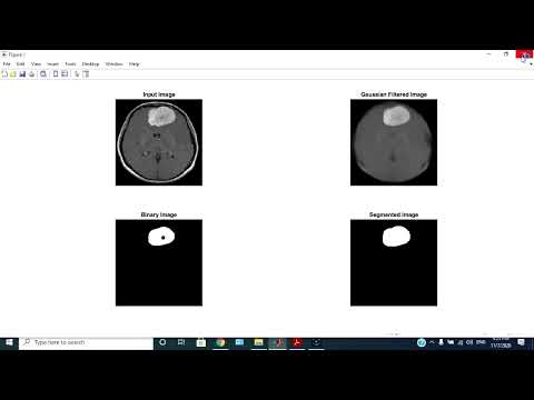
To detect infected tumor tissues from medical imaging modalities, segmentation is employed. Segmentation is necessary and important step in image analysis; it is a process of separating an image into different regions or blocks sharing common and identical properties, such as color, texture, contrast, brightness, boundaries, and gray level. Brain tumor segmentation involves the process of separating the tumor tissues such as edema and dead cells from normal brain tissues and solid tumors, such as WM, GM, and CSF with the help of MR images or other imaging modalities.
OBJECTIVE:
Brain tumor is the irregular and intense growth of tissues causing cancer. The most used technique to diagnose brain tumor is Magnetic Resonance Imaging (MRI). Precise information about the affected area is crucial for the appropriate treatment. The main objective of this paper is to develop an automated and appropriate method for detecting brain abnormalities and tumor edema using sobel edge detection method.
• The main aim of the application is tumor identification.
• The main reason behind the development of this application is to provide proper treatment as soon as possible and protect the human life which is in danger.
PROBLEM STATEMENT:
Irregular form and confusing boundaries of tumors make tumor segmentation more challenging.

• Demo Video
• Complete project
• Full project report
• Source code
• Complete project support by online
• Life time access
• Execution Guidelines
• Immediate (Download)
Software Requirements:
1. Matlab 2016A and Above
2. Image processing toolbox
Hardware Requirements:
1. PC or Laptop
2. 500GB HDD with 1 GB above RAM
3. Keyboard and mouse
1. Immediate Download Online