Introduction to Cervical Cancer Detection Using Deep Learning
Cervical cancer is a major healthcare concern that requires timely screening and accurate examination of cervical cell abnormalities. Early identification of abnormal cellular changes can support further clinical investigation and appropriate medical evaluation.
Traditional cervical cell examination involves analysing microscopic images to identify changes in cell structure, shape and appearance. This process can require significant time and specialist expertise.
Artificial intelligence and deep learning offer new opportunities to automate medical image analysis. Advanced convolutional neural networks can learn important visual patterns from cervical cell images and assist in identifying potentially abnormal cells.
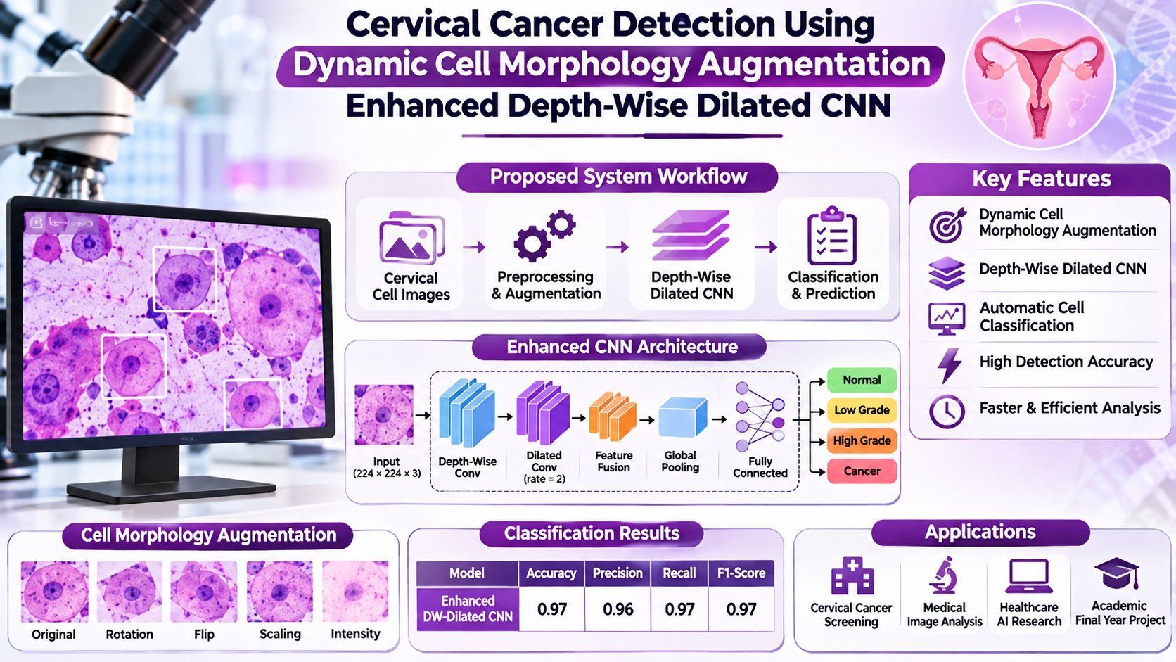
Cervical Cancer Detection Using Dynamic Cell Morphology Augmentation Enhanced Depth-Wise Dilated CNN is a deep learning-based project designed to analyse cervical cell images and classify cellular patterns using an enhanced CNN architecture.
The proposed approach combines dynamic cell morphology augmentation with depth-wise dilated convolution techniques to improve image feature extraction and classification performance.
Key Features & Benefits
Objectives of the Proposed System
The main objective is to develop an intelligent cervical cell image classification framework using advanced deep learning techniques.
Analyse microscopic cervical cell images.
Apply dynamic cell morphology augmentation to improve training data diversity.
Extract important cellular features such as shape, texture and structural patterns.
Develop an enhanced depth-wise dilated CNN architecture.
Classify cervical cell images into predefined categories.
Evaluate model performance using standard classification metrics.
Provide an automated interface for uploading and analysing images.
Implementation Guide
Technologies and Deep Learning Techniques
The proposed system can be implemented using Python-based computer vision and deep learning technologies.
Technologies Used:
Python for model development.
TensorFlow and Keras for deep learning.
OpenCV for image preprocessing.
NumPy and Pandas for data processing.
Matplotlib for visualization.
Scikit-learn for model evaluation.
Flask or Streamlit for application development.
Deep Learning Techniques:
Convolutional Neural Network (CNN): Extracts important visual features from cervical cell images for classification.
Dynamic Cell Morphology Augmentation: Applies suitable image transformations to increase training diversity while preserving relevant cellular structures.
Depth-Wise Convolution: Separates spatial filtering and channel-wise processing to make convolution operations more computationally efficient.
Dilated Convolution: Expands the receptive field of convolution filters, helping the model capture features at different spatial scales.
Enhanced CNN Architecture: Combines these techniques to learn meaningful cellular patterns and improve image classification.
Technical Specifications
Working Process of the Cervical Cancer Detection System
Step 1: Dataset Collection
Collect labelled cervical cell images from an appropriate medical image dataset.
Step 2: Image Preprocessing
Resize images, normalize pixel values and prepare the dataset for deep learning.
Step 3: Dynamic Morphology Augmentation
Apply suitable transformations such as rotation, flipping, scaling and other morphology-preserving augmentation techniques.
Step 4: Feature Extraction
The enhanced depth-wise dilated CNN extracts cellular features, including shape, texture and spatial characteristics.
Step 5: Image Classification
The trained model classifies the input image into the predefined categories available in the dataset.
Step 6: Performance Evaluation
Evaluate the trained model using accuracy, precision, recall, F1-score and confusion matrix.
Step 7: Result Visualization
Display the predicted classification and relevant analysis information through a simple web-based interface.
Combines image augmentation with advanced CNN techniques.
Extracts cellular features at different spatial scales.
Supports efficient deep learning experimentation.
Provides a foundation for AI-assisted screening tools.
Can be integrated into web-based medical image analysis applications.
Future Enhancements
The system can be extended with explainable AI, attention mechanisms, larger and more diverse datasets, cell-level segmentation and mobile-based image analysis. External validation using independent datasets can also help evaluate how well the model generalizes to different clinical image sources.
Conclusion
Deep learning has significant potential in medical image analysis and cervical cell classification. By combining dynamic cell morphology augmentation with depth-wise dilated CNN architecture, this project explores an advanced approach to automated cervical cell image analysis.
The proposed system can support research into AI-assisted cervical cancer screening. However, its predictions are not a medical diagnosis and must not replace professional screening, laboratory testing or evaluation by qualified healthcare professionals.
Contact Details
Aislyn Technologies, Bangalore
Phone: +91 97395 94609
Email: info@aislyntech.com
Website: https://aislyn.in
Contact us today to start building your Embedded project in Bangalore with our expert support!