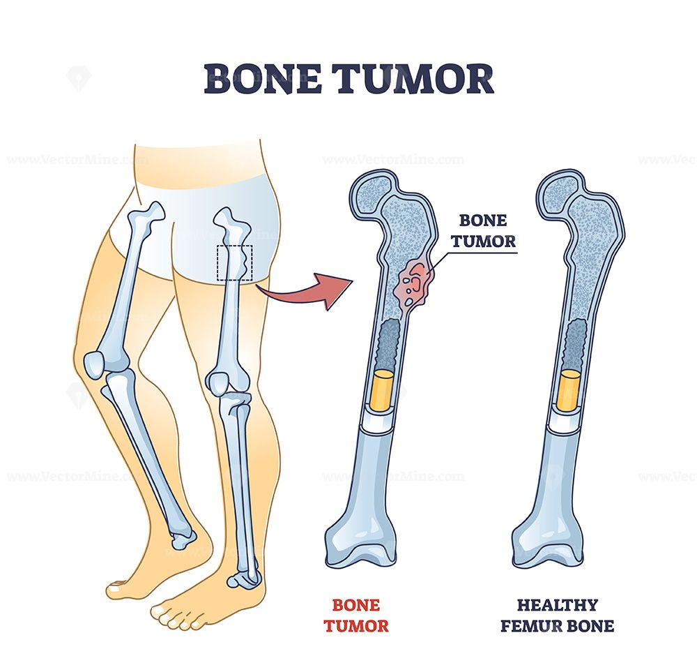
Bone tumours are abnormal growths that develop in or around bone tissue. Medical imaging techniques such as X-rays, CT scans, and MRI scans help healthcare professionals examine bone structures and identify abnormalities. Reviewing medical images can be challenging, particularly when large numbers of images need to be analyzed.
The Bone Tumour Detection System project explores the use of image processing and artificial intelligence to analyze medical images and identify patterns associated with bone abnormalities. By applying image preprocessing and deep learning techniques, the system can classify images into predefined categories based on its training dataset.
This project demonstrates how computer vision and deep learning can contribute to medical image analysis research. It is particularly useful for students interested in healthcare technology, image processing, and AI-based classification systems.
Key Features & Benefits
Working Methodology
The system begins with a collection of labelled bone images, such as X-ray images, organized according to the classes defined in the dataset. Before model training, images are preprocessed to improve consistency and prepare them for analysis.
Image preprocessing may include resizing, normalization, noise reduction, contrast enhancement, and data augmentation. These techniques help prepare images for the classification model.
A Convolutional Neural Network (CNN) can then extract important visual features from the images. The model learns patterns from the training dataset and uses the learned information to predict the category of an uploaded image.
Basic Workflow:
Collect and organize labelled bone image datasets.
Remove unsuitable or duplicate images.
Resize and preprocess the images.
Divide the dataset into training, validation, and testing sets.
Train the deep learning model using labelled images.
Evaluate the model with unseen test images.
Upload a new image through the application.
Display the predicted image category and confidence score.
Implementation Guide
Features and Technologies
Key Features
Medical image upload and analysis.
Image preprocessing and enhancement.
CNN-based image classification.
Automatic visual feature extraction.
Classification based on trained image categories.
Prediction confidence display.
Model performance evaluation.
User-friendly web interface.
Support for future dataset expansion.
Technologies Used
Programming Language: Python.
Deep Learning: TensorFlow and Keras.
Model Architecture: CNN.
Image Processing: OpenCV and Pillow.
Data Processing: NumPy and Pandas.
Visualization: Matplotlib.
Machine Learning Evaluation: Scikit-learn.
Backend: Flask or Streamlit.
Frontend: HTML, CSS and JavaScript.
Development Tools: Jupyter Notebook and VS Code.
Applications: Medical image analysis research, radiology education, computer vision projects, healthcare AI research, and academic deep learning demonstrations.
Technical Specifications
Implementation and Future Scope
The implementation begins with preparing a labelled bone image dataset. The images are divided into training, validation, and testing subsets to help measure how well the model performs on data it has not previously seen.
A CNN architecture is developed using convolutional layers for feature extraction, pooling layers for dimensionality reduction, and dense layers for classification. The model is trained using the prepared dataset, and its performance is evaluated using accuracy, precision, recall, F1-score, and a confusion matrix.
Once training is complete, the model can be integrated into a web application. Users upload a medical image, which is preprocessed before being passed to the trained model. The application displays the predicted category and associated confidence score.
Advantages
Automates image classification for research purposes.
Demonstrates practical medical image processing.
Learns visual features from labelled images.
Provides a simple image upload interface.
Supports repeatable model evaluation.
Combines computer vision and deep learning technologies.
Future Enhancements
Explore transfer learning using ResNet, DenseNet, or EfficientNet.
Expand the dataset with clinically verified images.
Integrate explainable AI techniques such as Grad-CAM.
Support additional medical imaging formats.
Develop a mobile-friendly application.
Improve model robustness across different imaging devices.
Evaluate performance using independent clinical datasets.
Conclusion & Next Steps
Conclusion and References
The Bone Tumour Detection System demonstrates how image processing and deep learning can be used to classify medical images into predefined categories. By preparing image datasets, extracting visual features, and training a CNN model, the project provides a practical example of AI-based medical image analysis.
This project is suitable for students interested in image processing, computer vision, Python programming, and deep learning. It offers practical experience in dataset preparation, model development, evaluation, and application integration.
Important Medical Disclaimer: This project is an academic research prototype and must not be used as a standalone diagnostic system. Its predictions do not confirm whether a person has a bone tumour. Medical diagnosis requires qualified healthcare professionals, appropriate imaging interpretation, and additional clinical investigations when necessary.