Introduction to Mycobacterium Tuberculosis Classification Using Fuzzy Logic
Tuberculosis (TB) is an infectious disease caused by the bacterium Mycobacterium tuberculosis. It primarily affects the lungs and can spread through the air when an infected person coughs or sneezes. Early identification and appropriate medical evaluation are important for controlling the spread of tuberculosis and supporting timely treatment.
Sputum microscopic examination is one of the established laboratory approaches used to identify acid-fast bacilli in patient samples. However, manually examining microscopic images requires trained laboratory personnel, careful observation, and considerable time, particularly when processing large numbers of samples.
The Classification and Counting of Mycobacterium Tuberculosis from Sputum Microscopic Image Using Fuzzy Logic project explores an automated image-processing approach for identifying and estimating bacterial objects in microscopic images.
The system uses image processing techniques to enhance microscopic images, identify candidate bacterial regions, and extract important visual characteristics. Fuzzy Logic is then applied to handle variations in image intensity, object shape, size, and other visual characteristics during classification.
Unlike conventional binary decision techniques, fuzzy logic uses degrees of membership to represent how strongly an object satisfies particular classification conditions. This makes it useful for investigating image classification problems where object boundaries and visual characteristics may not always be clearly defined.
This Python-based project is suitable for students interested in Medical Image Processing, Artificial Intelligence, Fuzzy Logic, Computer Vision, and automated microscopic image analysis.
Key Features & Benefits
Project Concept and Working Methodology
The primary objective of this project is to develop an automated system for classifying and counting Mycobacterium tuberculosis candidates from sputum microscopic images using image processing and fuzzy logic techniques.
The proposed system follows a sequence of image-processing and classification operations.
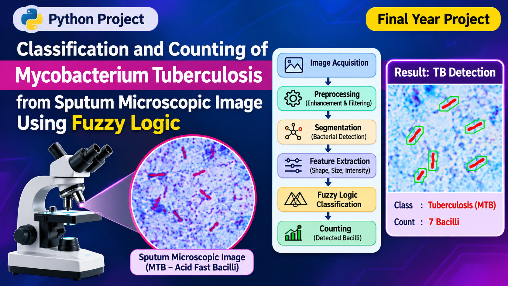
1. Microscopic Image Acquisition: Sputum microscopic images are collected from an appropriately labeled dataset or compatible digital microscopy source.
2. Image Preprocessing: The input images are converted into a suitable color or grayscale representation to simplify subsequent processing.
3. Image Enhancement: Image enhancement techniques such as contrast adjustment, noise reduction, and filtering are applied to improve the visibility of candidate bacterial structures.
4. Image Segmentation: Thresholding and morphological operations are used to separate potential bacterial objects from the surrounding microscopic background.
5. Feature Extraction: Important visual characteristics such as object area, shape, aspect ratio, intensity, and color information are extracted from the segmented regions.
6. Fuzzy Membership Calculation: Fuzzy membership functions are defined for selected features. These functions represent the degree to which an identified object satisfies different classification conditions.
7. Fuzzy Rule Evaluation: A fuzzy inference system applies predefined IF–THEN rules to evaluate the extracted features and determine the classification of candidate objects.
8. Object Classification: Candidate regions are classified according to their fuzzy membership scores and the selected decision rules.
9. Bacterial Counting: Objects satisfying the configured classification criteria are counted to generate an estimated bacterial count for the input image.
10. Result Visualization: The application displays the processed microscopic image, highlighted candidate regions, classification information, and estimated object count.
This methodology combines image enhancement, segmentation, feature extraction, and fuzzy inference to investigate automated microscopic image analysis.
Implementation Guide
Key Features and Real-World Applications
The proposed fuzzy logic-based microscopic image analysis system provides several features for studying bacterial object classification and counting.
Key Features:
Sputum microscopic image uploading.
Automated image preprocessing.
Microscopic image enhancement.
Noise reduction and filtering.
Bacterial candidate region segmentation.
Visual feature extraction.
Fuzzy membership function calculation.
Rule-based fuzzy classification.
Candidate object identification.
Automated estimated object counting.
Highlighted classification results.
Processed image visualization.
Graphical display of analysis results.
Real-World Applications:
Medical Image Processing Research: Provides a framework for studying automated analysis of microscopic medical images.
Tuberculosis Research: Supports experimental investigation of image-processing methods for identifying acid-fast bacilli candidates.
Laboratory Image Analysis: Demonstrates how digital microscopy images can be processed using computer vision techniques.
Automated Microscopy Research: Provides a foundation for developing intelligent microscopic image analysis applications.
Academic Research: Helps students understand fuzzy inference systems, image segmentation, feature extraction, and classification.
Medical AI Development: Can be extended into research involving other microscopic image classification tasks.
The system is intended for experimental image analysis. Its outputs should not be considered confirmed tuberculosis diagnoses or substitutes for validated laboratory testing and professional medical evaluation.
Technical Specifications
Technologies and Software Requirements
The project can be developed using Python and image-processing libraries combined with fuzzy logic techniques.
Development Environment: Visual Studio Code, Jupyter Notebook, or PyCharm.
Hardware Requirements:
Processor: Intel Core i3 or higher.
RAM: Minimum 8 GB recommended.
Storage: Minimum 5 GB available space.
Operating System: Windows 10/11 or Linux.
Graphics: Integrated GPU or compatible NVIDIA GPU.
Display: Standard monitor.
Input Devices: Keyboard and mouse.
Image Acquisition: Digital microscope or existing microscopic image dataset.
Fuzzy Logic Implementation:
Fuzzy logic is an important component of the proposed classification process. Instead of depending entirely on fixed thresholds, the system can use membership functions to represent different degrees of feature similarity.
For example, extracted object characteristics can be evaluated using fuzzy categories such as:
Small, medium, and large object size.
Low, medium, and high image intensity.
Low, medium, and high aspect ratio.
Weak, moderate, and strong candidate membership.
A fuzzy inference system combines these conditions using predefined rules to calculate an output classification score.
The extracted objects can then be evaluated against a selected decision threshold to estimate the number of candidate bacterial structures.
Conclusion & Next Steps
Advantages, Future Scope, and Conclusion
Advantages of the Project
The proposed system demonstrates how image processing and fuzzy logic can be combined for microscopic image analysis.
Reduces dependence on manual object counting during experimental analysis.
Supports feature-based candidate classification.
Uses fuzzy membership values to handle uncertain visual characteristics.
Provides visual identification of candidate regions.
Generates estimated object counts.
Supports repeatable image-processing workflows.
Provides a foundation for advanced medical image analysis research.
Future Enhancements
The project can be extended with modern Artificial Intelligence and Computer Vision technologies.
Integration of CNN-based bacterial image classification.
Development of deep learning-based object detection.
Integration of YOLO for candidate object localization.
Improved segmentation using advanced thresholding techniques.
Comparison of fuzzy logic with traditional machine learning classifiers.
Development of a real-time digital microscopy interface.
Automated image quality assessment.
Integration of explainable AI for visual classification analysis.
Cloud-based microscopic image storage and processing.
Development of a laboratory image analysis dashboard.
Future versions can also investigate hybrid neuro-fuzzy systems that combine neural network feature extraction with fuzzy inference for more advanced classification research.
Conclusion
Classification and Counting of Mycobacterium Tuberculosis from Sputum Microscopic Image Using Fuzzy Logic is an interesting Python-based project that combines image processing, feature extraction, fuzzy inference, and automated object counting.
By processing sputum microscopic images and evaluating candidate bacterial regions using fuzzy membership functions, the system demonstrates an approach to experimental microscopic image classification.
The project provides students with practical knowledge of Python programming, OpenCV, fuzzy logic, image segmentation, feature extraction, and computer vision. It also establishes a foundation for exploring advanced AI-based medical image analysis and automated microscopy research.
The developed system should be independently validated using appropriately labeled clinical datasets and expert-reviewed results before any diagnostic application is considered.
Contact Aislyn Technologies – Bangalore
Aislyn Technologies
📞 Phone: +91 97395 94609
📧 Email: info@aislyntech.com
🌐 Website: https://aislyn.in
Looking to develop a Python, Artificial Intelligence, Machine Learning, Medical Image Processing, or Deep Learning project? Contact Aislyn Technologies for expert project development guidance, implementation support, and customized final-year project solutions in Bangalore.

























.png)


