Introduction to Automated Left Atrial Segmentation
The human heart plays an essential role in circulating blood throughout the body. Understanding its structure and function is important for cardiovascular research and medical assessment. Magnetic Resonance Imaging (MRI) provides detailed images of cardiac structures and helps researchers examine the heart from different perspectives.
The left atrium is an important chamber that receives oxygen-rich blood from the lungs. Identifying its boundaries across multiple MRI images can help researchers study cardiac anatomy and volume changes.
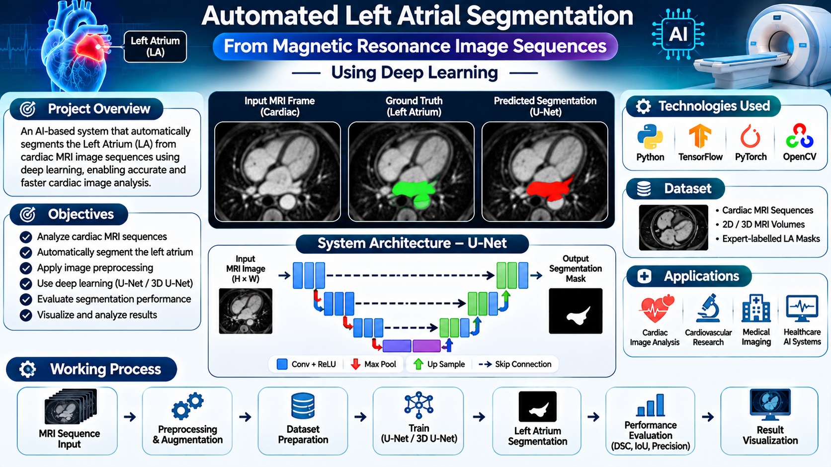
Automated Left Atrial Segmentation from Magnetic Resonance Image Sequences is a machine learning project designed to identify and separate the left atrium from cardiac MRI images. The system uses medical image preprocessing and deep learning techniques to generate segmentation masks with reduced manual intervention.
This approach explores how artificial intelligence can support cardiac image analysis, anatomical visualization and cardiovascular research.
Key Features & Benefits
Objectives of the Proposed System
Analyse cardiac MRI image sequences.
Automatically identify the left atrial region.
Apply image preprocessing for improved segmentation.
Extract important anatomical features from MRI images.
Generate pixel-level segmentation masks.
Reduce repetitive manual image annotation.
Evaluate segmentation performance using standard metrics.
Visualize segmented cardiac structures for further analysis.
Implementation Guide
Technologies and Deep Learning Algorithms
Technologies Used:
Python: Main programming language.
TensorFlow or PyTorch: Deep learning model development.
OpenCV: Medical image preprocessing and visualization.
NumPy: Numerical image operations.
Pandas: Dataset organization.
Scikit-learn: Model evaluation.
Matplotlib: Visualization of MRI images and segmentation results.
Flask or Streamlit: Web-based application interface.
Deep Learning Algorithms
U-Net: A popular encoder-decoder architecture for biomedical image segmentation that combines contextual information with spatial details.
3D U-Net: Processes volumetric MRI data and learns spatial relationships across multiple image slices.
Convolutional Neural Network (CNN): Extracts anatomical features from cardiac MRI images.
Attention U-Net: Uses attention mechanisms to emphasize relevant anatomical regions during segmentation.
Dice Loss: Helps optimize segmentation models by measuring overlap between predicted and ground-truth regions.
Evaluation Metrics
The model can be evaluated using Dice Similarity Coefficient (DSC), Intersection over Union (IoU), precision, recall and Hausdorff distance.
Technical Specifications
Working Process of the Left Atrial Segmentation System
Step 1: MRI Dataset Collection
Collect cardiac MRI sequences with corresponding expert-labelled segmentation masks.
Step 2: Image Preprocessing
Prepare MRI images through resizing, normalization and other suitable preprocessing operations.
Step 3: Dataset Preparation
Organize the images and their corresponding masks into training, validation and testing datasets.
Step 4: Deep Learning Model Training
Train a selected segmentation architecture such as U-Net or 3D U-Net using labelled MRI images.
Step 5: Feature Extraction
The network learns important anatomical patterns and spatial characteristics of the left atrium.
Step 6: Segmentation Prediction
Provide an MRI image or sequence to the trained model to generate a predicted left atrial segmentation mask.
Step 7: Result Visualization
Display the original MRI image alongside the predicted segmentation overlay for visual inspection.
Step 8: Performance Evaluation
Compare predicted masks with expert-labelled masks using segmentation evaluation metrics.
Conclusion & Next Steps
Applications and Advantages
Applications
Cardiac MRI image analysis.
Cardiovascular research.
Left atrial anatomical visualization.
Medical image segmentation research.
AI-based healthcare applications.
Academic machine learning projects.
Advantages
Automates left atrial image segmentation.
Reduces repetitive manual annotation work.
Supports analysis of multiple MRI slices.
Provides visual segmentation outputs.
Enables experimentation with advanced deep learning models.
Supports quantitative evaluation of cardiac structures.
Future Enhancements
The project can be enhanced with attention-based segmentation, 3D visualization, multi-structure cardiac segmentation, explainable AI and integration with medical imaging platforms. Testing on independent MRI datasets can also help evaluate model generalization across different scanners and imaging protocols.
Conclusion
Automated left atrial segmentation demonstrates the potential of deep learning in cardiovascular medical image analysis. By combining cardiac MRI preprocessing, U-Net-based architectures and segmentation evaluation techniques, this project provides a foundation for automated anatomical structure identification.
The system can support medical imaging research and image analysis workflows. Its outputs should be reviewed by qualified healthcare professionals and should not be treated as a standalone diagnostic result.
Contact Details
Aislyn Technologies, Bangalore
Phone: +91 97395 94609
Email: info@aislyntech.com
Website: https://aislyn.in
Contact us today to start building your Embedded project in Bangalore with our expert support!