Blood analysis plays an important role in identifying different blood-related conditions and supporting medical investigations. Traditional blood testing often involves laboratory procedures and manual examination by trained professionals. With the development of artificial intelligence, image processing, and machine learning, researchers are exploring computer-based approaches to analyze microscopic blood images and support laboratory workflows.
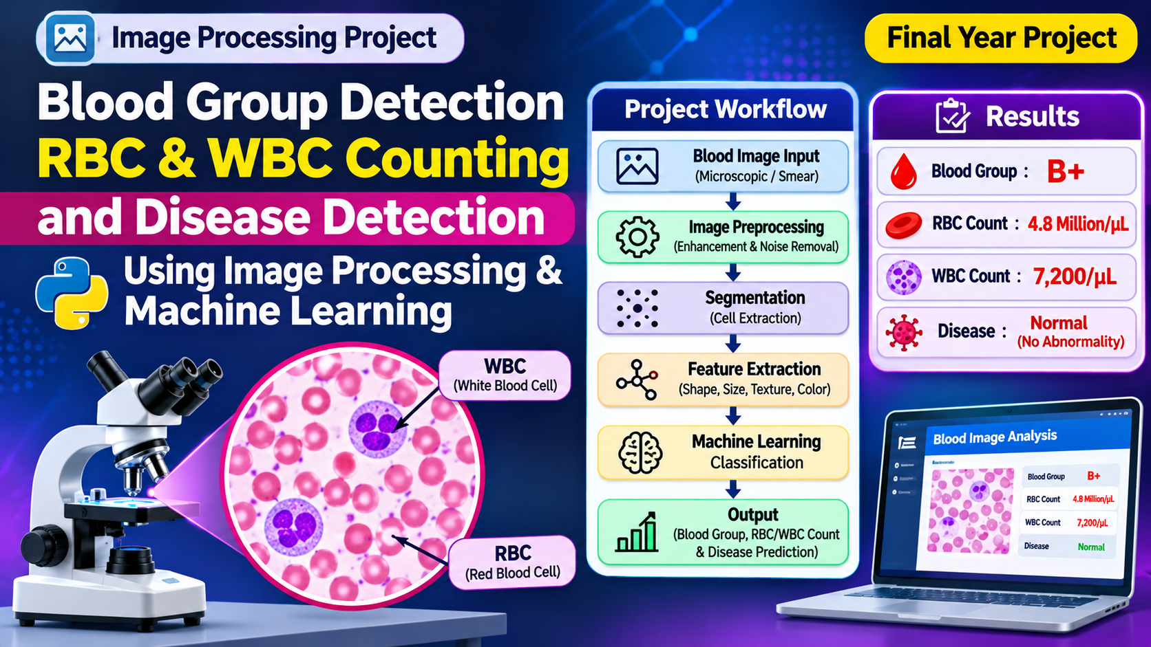
The Blood Group Detection, RBC WBC Counting and Disease Detection Using an Image Processing Approach and Machine Learning project focuses on analyzing blood-related images through digital image processing techniques. It explores the identification of blood group characteristics, counting red blood cells (RBCs) and white blood cells (WBCs), and identifying selected abnormal patterns that may be associated with blood-related diseases.
The system can process input images, improve image quality, separate relevant cellular regions, and extract useful features for further analysis. Machine learning techniques can then be applied to classify image patterns based on the dataset used for training.
This project demonstrates how image processing and machine learning can be combined to develop an intelligent blood image analysis prototype for academic research and educational purposes.
Key Features & Benefits
Project Concept and Working Methodology
The main concept of this project is to convert microscopic blood images into useful digital information through image processing and machine learning.
The workflow begins with uploading or capturing a blood sample image. The image is then processed to improve its quality and prepare it for analysis. Depending on the image quality and staining method, preprocessing may include resizing, noise removal, contrast enhancement, and colour-space conversion.
After preprocessing, segmentation techniques are used to separate blood cells from the background. The system can identify regions that potentially represent red blood cells and white blood cells. Image features such as shape, size, colour, and texture can be extracted to support cell classification and counting.
A machine learning model can use labelled training images to learn patterns associated with the selected blood image categories. Once trained, the model can analyze new images and produce predicted classifications.
Typical project workflow:
Blood image acquisition – Collect microscopic blood images from a suitable dataset or laboratory imaging source.
Image preprocessing – Resize images, reduce noise, and enhance contrast.
Image segmentation – Separate relevant cell regions from the background.
RBC and WBC identification – Detect and classify cell regions using image-processing or trained machine learning methods.
Cell counting – Estimate the number of identified cells in the processed image.
Blood group classification – Apply a suitable image-based classification method if the required labelled dataset and validated methodology are available.
Disease-pattern analysis – Examine selected image characteristics and classify patterns represented in the training dataset.
Result presentation – Display the analysis results through a user interface.
The actual performance depends on image quality, dataset diversity, annotation accuracy, model selection, and experimental validation.
Implementation Guide
Key Features and Applications
The project combines multiple image analysis concepts into a single research-oriented application.
Key Features
Blood Image Upload: Allows users to provide microscopic blood images for processing.
Image Preprocessing: Improves image quality through filtering, resizing, and contrast enhancement.
RBC Detection: Identifies red blood cell regions in microscopic images.
WBC Detection: Identifies white blood cell regions based on visual characteristics.
RBC and WBC Counting: Estimates cell counts from detected image regions.
Blood Group Classification: Provides an experimental classification module when supported by an appropriate blood-group-labelled dataset and validated testing procedure.
Disease Pattern Analysis: Examines selected image features and predicts categories learned from the training data.
Result Visualization: Presents processed images, detected regions, and model predictions.
Machine Learning Integration: Uses trained models to support image classification and pattern recognition.
Potential Applications
Academic research in medical image processing.
Educational demonstrations of blood cell segmentation and classification.
Experimental automated microscopy workflows.
Machine learning research using labelled blood image datasets.
Development of prototype computer-assisted laboratory tools.
An important distinction is that RBC/WBC morphology analysis does not independently establish a patient's diagnosis. Likewise, blood group determination generally requires validated serological testing, such as antigen and antibody-based testing; ordinary microscopic image classification should not be treated as a replacement.
Technical Specifications
Technologies, Software and Hardware Requirements
The project can be implemented using a combination of programming, image processing, machine learning, and database technologies. The final technology selection depends on the implementation.
Software Requirements
Programming Language: Python
Image Processing: OpenCV
Machine Learning: Scikit-learn
Deep Learning: TensorFlow or Keras
Numerical Processing: NumPy
Data Analysis: Pandas
Visualization: Matplotlib
User Interface: Flask or Streamlit
Development Environment: Visual Studio Code, PyCharm, or Jupyter Notebook
Database: SQLite or MySQL, if result storage is required
Operating System: Windows or Linux
Hardware Requirements
Processor: Intel Core i3 or above
RAM: Minimum 8 GB recommended
Storage: 10 GB free space or more
Display: Standard HD monitor
Camera or Microscope: Digital microscope or suitable image acquisition device
GPU: Optional, depending on the selected deep learning model
Machine Learning and Image Processing Techniques
Image enhancement, thresholding, morphological operations, contour detection, and segmentation can be used to identify cell regions. Feature extraction can capture visual properties such as shape, size, and texture. Depending on the dataset, classification may use traditional machine learning algorithms or convolutional neural networks (CNNs).
For reliable evaluation, the model should be tested on images that were not used during training. Performance can be measured using suitable metrics such as precision, recall, F1-score, and confusion matrices. Cell counting should also be evaluated against manually annotated reference images.
Conclusion & Next Steps
Advantages, Future Scope and Conclusion
Advantages
Demonstrates the practical combination of image processing and machine learning.
Reduces the amount of repetitive manual image examination in an experimental workflow.
Provides a structured approach to identifying and counting blood cell regions.
Supports visualization of processed microscopic images.
Offers opportunities to study segmentation, feature extraction, and image classification.
Can be extended with additional labelled datasets and machine learning models.
Future Scope
The project can be improved by training advanced deep learning models on larger and more diverse microscopic blood image datasets. Object detection and instance segmentation models may improve the localization and counting of overlapping cells. Additional features could include automated report generation, comparison of model predictions with expert annotations, and integration with laboratory information systems.
Further research could also investigate the effect of image quality, staining variations, microscope types, and dataset imbalance on model performance. Any clinical application would require appropriate validation, regulatory review, and evaluation by qualified medical professionals.
Conclusion
Blood Group Detection, RBC WBC Counting and Disease Detection Using Image Processing and Machine Learning is an interesting project that demonstrates the potential of artificial intelligence in microscopic blood image analysis. By combining preprocessing, segmentation, feature extraction, and machine learning, the system can provide an experimental platform for cell identification, counting, and image-based classification.
The project is suitable for students interested in image processing, artificial intelligence, machine learning, and healthcare-related research. Its outputs should be considered research predictions rather than confirmed medical results. Blood grouping and disease diagnosis must always be performed using validated laboratory procedures and reviewed by qualified healthcare professionals.