ABSTRACT
Brain tumor identification and segmentation from magnetic resonance imaging (MRI) scans remain challenging tasks in medical image analysis due to the complex structure of brain tissues and the variability in tumor appearance. Accurate and early detection is essential for effective diagnosis, treatment planning, and survival assessment of patients. Conventional manual analysis performed by radiologists is time-consuming and susceptible to subjective interpretation, which increases the demand for automated and reliable computational solutions. This project presents an intelligent brain tumor detection and segmentation system based on a deep learning–driven Residual U-Net (ResUNet) architecture. The proposed approach combines the advantages of encoder–decoder networks with residual learning to enhance feature extraction, improve gradient flow, and achieve precise localization of tumor regions. MRI images are preprocessed through resizing, normalization, and binary mask generation to ensure consistent training data quality. The ResUNet model is trained in a supervised manner using paired MRI images and ground-truth tumor masks, optimized with binary cross-entropy loss and adaptive learning rate strategies. Experimental evaluation demonstrates stable convergence and effective segmentation performance across validation samples. In addition to model development, a user-friendly graphical interface is designed using the Tkinter framework to facilitate real-time interaction. The interface allows users to upload MRI images, visualize original and processed outputs, and obtain segmentation results in an intuitive format. Tumor severity is estimated by calculating the segmented pixel area, enabling classification into different tumor size categories. Based on the detected tumor region, the system also provides an approximate survival timeline to assist preliminary clinical interpretation. The integration of deep learning-based segmentation with an interactive visualization platform enhances the practicality of the proposed system. Overall, this project highlights the potential of ResUNet-based models in automated brain tumor analysis and demonstrates their applicability as a supportive tool in medical imaging and decision-assistance systems.
INTRODUCTION
Brain tumors represent one of the most critical and life-threatening neurological disorders due to their direct impact on cognitive, sensory, and motor functions of the human brain. A brain tumor is characterized by the abnormal and uncontrolled growth of cells within the brain or surrounding tissues, which can disrupt normal neural activity and lead to severe health complications. Magnetic Resonance Imaging (MRI) is widely regarded as the most effective imaging modality for brain tumor diagnosis because of its high spatial resolution and superior contrast between soft tissues. Despite the availability of advanced imaging techniques, accurate interpretation of MRI scans remains a complex and time-consuming task that requires significant clinical expertise. Manual tumor identification and segmentation performed by radiologists are often subject to inter-observer variability, fatigue-related errors, and delays, particularly when handling large volumes of patient data. These limitations highlight the urgent need for automated, efficient, and reliable computer-aided diagnostic systems.
Recent advancements in artificial intelligence and deep learning have significantly transformed the field of medical image analysis by enabling automated feature learning and high-precision image segmentation. Convolutional Neural Networks (CNNs) have emerged as a dominant approach for extracting hierarchical features from medical images without the need for handcrafted descriptors. Among CNN-based architectures, encoder–decoder models such as U-Net have demonstrated exceptional performance in biomedical segmentation tasks. However, conventional U-Net architectures may encounter challenges such as vanishing gradients and insufficient feature reuse when trained on deep networks with complex tumor boundaries. To address these issues, residual learning mechanisms have been integrated into segmentation frameworks, giving rise to the Residual U-Net (ResUNet) architecture. The ResUNet model enhances the learning capability of the network by introducing skip connections within convolutional blocks, allowing the model to learn residual mappings rather than direct transformations. This design improves gradient propagation during training and enables the extraction of more discriminative features from MRI images. In this project, a ResUNet-based segmentation model is developed to automatically identify and segment tumor regions from brain MRI scans with high accuracy. The system is trained using a curated dataset consisting of MRI images and their corresponding ground-truth tumor masks. Preprocessing steps such as image resizing, normalization, and binary mask thresholding are employed to standardize the dataset and improve model generalization.
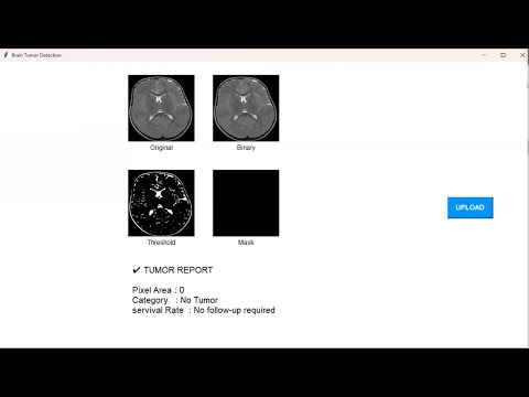
In addition to the deep learning model, this project emphasizes practical applicability by integrating the trained ResUNet model into a graphical user interface developed using the Tkinter framework. The interface allows users to upload MRI images, visualize intermediate processing stages, and obtain tumor segmentation results in real time. To provide clinically relevant insights, the segmented tumor area is analyzed to estimate tumor size and categorize severity levels. Based on the computed tumor region, an approximate survival timeline is presented to assist in preliminary clinical assessment. By combining automated segmentation, visual interpretation, and user interaction, the proposed system serves as a comprehensive decision-support tool. Overall, this project demonstrates the effectiveness of deep learning-based ResUNet architectures in brain tumor detection and highlights their potential role in improving diagnostic efficiency, consistency, and accessibility in modern healthcare systems.
The increasing availability of large-scale medical imaging data has further accelerated the adoption of deep learning techniques in healthcare applications. Brain MRI datasets often contain complex variations in tumor size, shape, intensity, and location, making automated analysis a challenging task. Tumors may appear with irregular boundaries and heterogeneous textures, which complicates traditional image processing and threshold-based segmentation methods. Deep learning models, particularly convolutional neural networks, address these challenges by learning robust feature representations directly from data, thereby reducing dependency on handcrafted rules and domain-specific heuristics. As a result, automated tumor segmentation systems can achieve higher consistency and repeatability compared to manual approaches. Another significant challenge in brain tumor analysis is the differentiation between tumor and normal brain tissues, as well as the accurate estimation of tumor extent. Even small segmentation inaccuracies can lead to incorrect tumor grading and may adversely affect treatment planning, including surgical resection and radiotherapy dosage. The incorporation of residual connections within deep segmentation networks enables the model to capture both low-level spatial details and high-level semantic information. This balance is crucial for preserving fine tumor boundaries while maintaining contextual understanding of surrounding brain structures. The ResUNet architecture employed in this project is specifically designed to address these requirements by combining residual learning with multi-scale feature fusion.

• Demo Video
• Complete project
• Full project report
• Source code
• Complete project support by online
• Life time access
• Execution Guidelines
• Immediate (Download)
SYSTEM REQUIREMENTS
Hardware Requirements
1. Intel Core i5 processor or equivalent to support image processing and deep learning computations.
2. Multi-core CPU recommended for faster training and efficient multitasking.
3. Minimum 8 GB RAM to handle MRI datasets and intermediate tensor operations.
4. Dedicated GPU (NVIDIA GTX/RTX with CUDA support) recommended for accelerated model training.
5. At least 50 GB of free storage for dataset files, trained models, and result outputs.
6. Standard display unit with a minimum resolution of 1366 × 768 for clear visualization of images.
7. Keyboard and mouse for user interaction with the graphical interface.
Software Requirements
1. Operating System: Windows / Linux / macOS (64-bit).
2. Python version 3.8 or higher for model development and execution.
3. TensorFlow and Keras libraries for building and training the ResUNet model.
4. OpenCV library for image processing and mask handling.
5. NumPy library for numerical computation and array manipulation.
6. Matplotlib library for plotting training accuracy and loss graphs.
7. Tkinter library for developing the graphical user interface.
8. Pillow (PIL) library for image rendering within the GUI.
9. Integrated Development Environment (IDE) such as PyCharm or Visual Studio Code.
1. Immediate Download Online